Air leaks in lungs
| Summary | |
|---|---|
| Date | 2021-2024 |
| Tags | full field measurements, digital image correlation, composite materials, non local modeling |
| Partners | Pr. Isabelle Villemure, Dr. George Rakovich M.D. |
| Recruiting | Magalie Mambangui |
First phase of the project
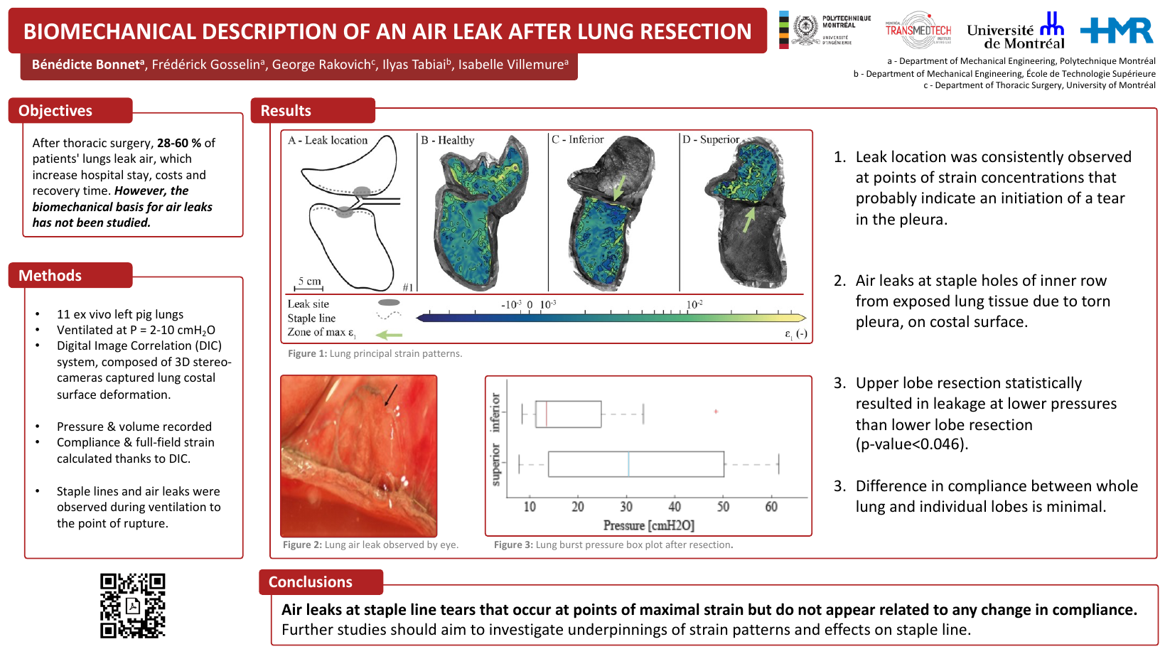
The first phase of this project was carried out by Bénédicte Bonnet (now a happy Msc in biomedical engineering) and mainly aimed at developing an experimental setup that can be used to quantify and better understand the presence of air leaks in lungs after resection. The study was supervised by Pr. Frédérick Gosselin, Pr. Isabelle Villemure and Dr. George Rakovich M.D.. There will be a publication for this one, it is currently under review.
A pair of pork lungs donated to science are connected to a piston that is controlled to inflate and deflate them. A stereoscopic DIC setup (white paint base with a speckle pattern on the lung, two cameras and lights) is used to capture consecutive stereo-images that are used for DIC analysis. The animation on the right presents a contour plot showing the evolution of the strain in the principal direction on the lung’s surface.
The animation on the right shows that lungs do not deform homogeneously during inflation, some areas are much more solicited that others, while some areas barely deform. The complete study aimed at studying the impact of lung resection (cutting and removing a section of lung) that is often sealed using a special staple gun. DIC was in this case able to reveal the impact of stapling lung tissue close to the staples and further away from them.
The work of Bénédicte Bonnet is summarized in the following poster:
Second phase of the project
Pr. Isabelle Villemure, Dr. George Rakovich M.D. and myself are currently pursuing this project with a second phase that aims at studying the local mechanics and behavior of airleaks that appear after resection. We are currently looking for an industrial partner in order to pursue this project.
Leave a comment